Melissa Venator studies an x-radiograph for George Grosz's Café on a lightbox.

When German physics professor Wilhelm Conrad Röntgen discovered x-radiographs in 1895, he captured the image of bones inside a human hand. More than 100 years later, art professionals at the Saint Louis Art Museum are using x-rays to uncover the brushstrokes of painted hands and more by examining layers hidden under the surface of an easel painting.
Since the 19th century, the core method of radiography remains unchanged: an energy source emits x-rays through an object placed in front of sensitive film. The film is processed digitally or chemically to produce the radiograph image. Materials transparent to x-rays appear dark in the radiograph, while denser, x-ray–absorbing materials appear white. In medical radiography, skin and tissue become transparent, allowing viewers to see through to the gleaming, sharply contrasted white bones. The same technique can be applied to easel paintings.
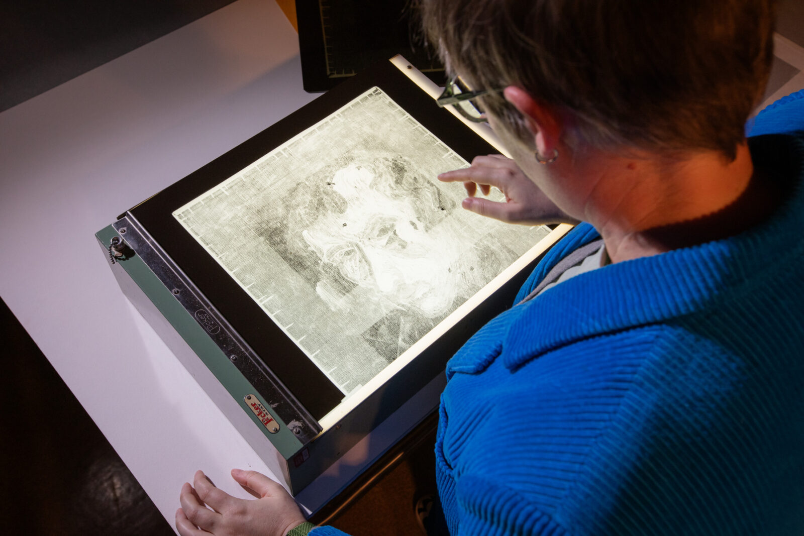
Melissa Venator studies an x-radiograph for George Grosz's Café on a lightbox.
Anatomy of a painting
The material “bones” of a painting, made of paint, canvas, and wooden frame (called a stretcher or strainer), are all visible in a radiograph. The intensity and clarity of each material in the image is determined by their ability to absorb x-rays.
All materials absorb x-rays to some degree; however, metals absorb x-rays extremely well. Paints that are rich with metallic elements, such as lead white, appear bright white in a radiograph. Paints colored with organic compounds, such as alizarin red and indigo blue, become transparent. Even though the resulting radiograph includes all material layers of the painting in a composite image, the push and pull of opacity and transparency render only some materials visible. Changes made by the artist—and, in some exciting instances, made completely different from what is visible to the naked eye—may be unveiled.

Franz Marc, German, 1880–1916; The Little Mountain Goats, 1913–14; oil on canvas; 23 7/8 x 16 inches; Saint Louis Art Museum, Bequest of Morton D. May 912:1983
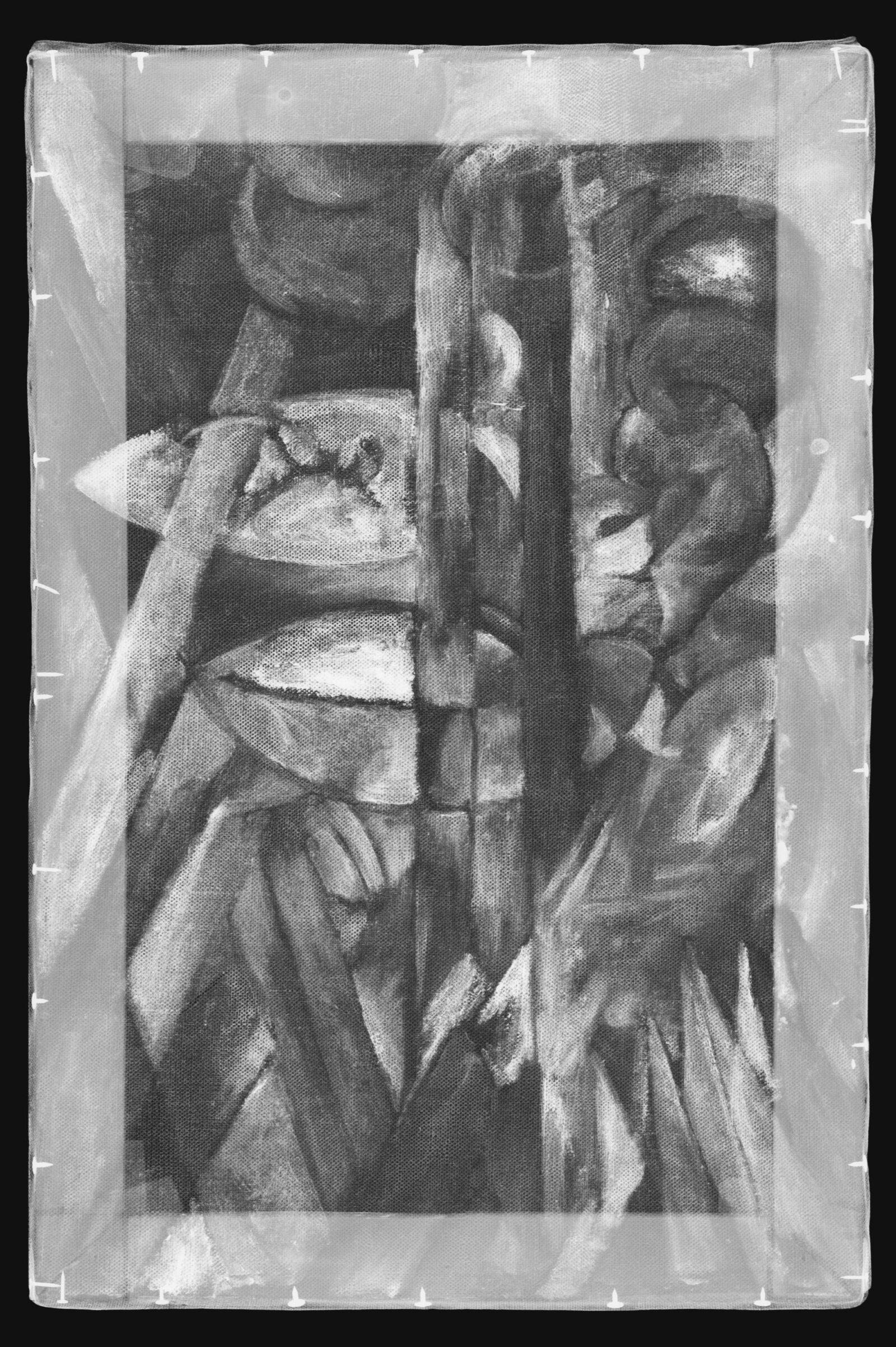
X-radiograph view of The Little Mountain Goats
Take for example The Little Mountain Goats, painted by Franz Marc in 1914. The painting is executed in oil paint on canvas, which is pulled taut over a wooden stretcher and fixed with nails. In the radiograph, the outline of the wooden stretcher is clearly visible, and the metal tacks securing the canvas to the stretcher appear bright white.
Looking closer, stretcher keys, small wooden wedges used to tension the stretched canvas, are also visible at the corners. Excess canvas is folded to align with the stretcher bars, and if one zooms in enough, the weave pattern of the fabric appears. Such details enable conservators to identify the materials of the painting and also diagnose condition issues, such as paint cracks or canvas tears.
Exploring the artist’s technique
Zooming out to view the full x-radiograph, paint layers that appear the most opaque bright white correspond to areas of metal-dense zinc white. The composition in the radiograph aligns with Marc’s final composition, confirming that the artist has stayed true to his initial design.

August Macke, German, 1887–1914; Landscape with Cows, Sailboat, and Painted-in Figures, 1914; oil on canvas; 20 1/4 × 20 1/4 inches; Saint Louis Art Museum, Bequest of Morton D. May 911:1983
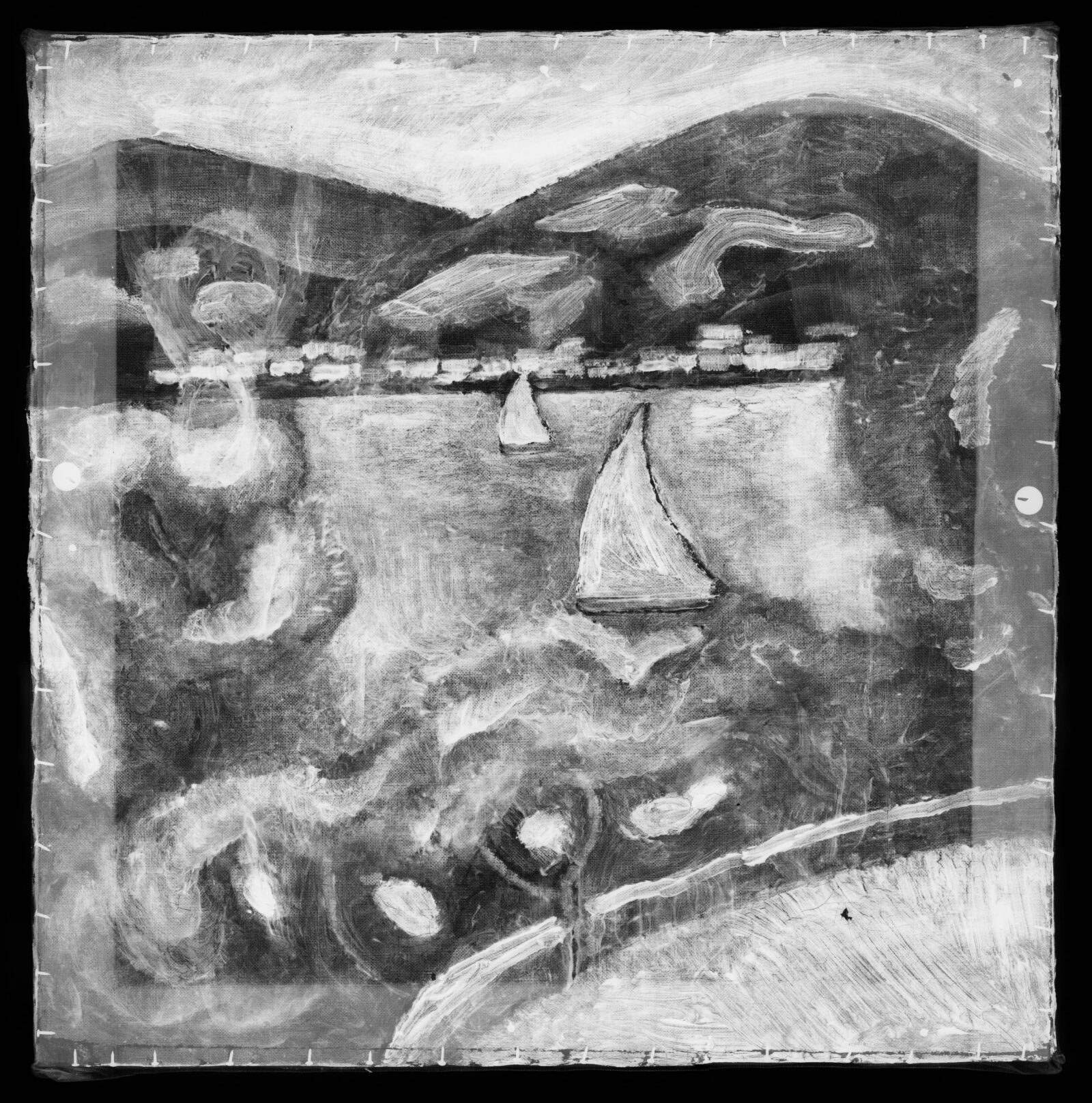
X-radiograph view of Landscape with Cows, Sailboat, and Painted-in Figures
Auguste Macke’s Landscape with Cows, Sailboat, and Painted-in Figures, dated 1914, offers a more revealing view into the artist’s creative process. Light cast from a sharp side angle onto the surface of the painting reveals paint textures that differ from the final image. This is a primary clue that the brushwork of other compositions may be lying underneath.
Radiography confirmed suspicions that the sailboats in the center belong to an underlying lake scene. Underneath the later paint additions of rotated figures, buildings, and animals is a fully rendered composition: a clear landscape with rolling mountains, a lake, a distant shoreside town, and boats sailing beyond a terrace in the foreground. A few years earlier, Macke painted very similar scenes depicting the Tegernsee, an alpine lake in Southern Germany. Why Macke made the rotating additions to the underlying painting in Landscape with Cows, Sailboat, and Painted-in Figures remains a mystery, but the painting’s uniqueness continues to intrigue viewers.
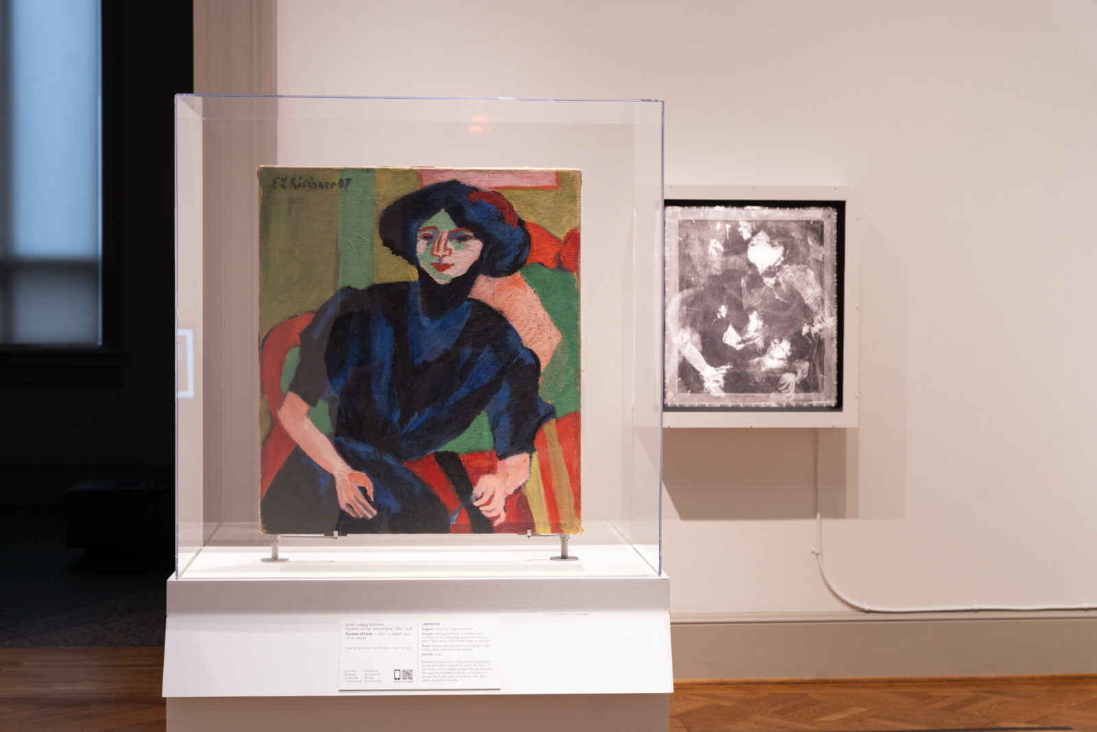
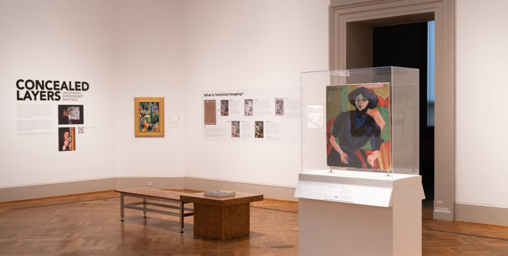
Installation view of Concealed Layers: Uncovering Expressionist Paintings featuring a painting and related x-radiograph in a lightbox
Through x-rays, conservators gain a wealth of stories told through a painting’s revealed materials and layers. Much like how a doctor may learn a broken bone is the result of a daring climb, a conservator can team with an art historian to study how an artist’s known technique or favored subject may explain an anomaly in a painting’s layers.
Explore radiographs set within bright lightboxes up close in Concealed Layers: Uncovering Expressionist Paintings. The exhibition presents recent discoveries from a comprehensive study of SLAM’s world-class collection of German Expressionist paintings. The exhibition is currently on view in Caro Nichols Holmes Gallery 214 and Sherry and Gary Wolff Gallery 215 through October 27.
Safety disclaimer: X-radiography is a nondestructive technique used in art conservation—the painting’s materials remain unaffected by the x-ray exposure. Proper health and safety procedures are taken to ensure that all Museum personnel are safe during the x-radiography of the paintings.