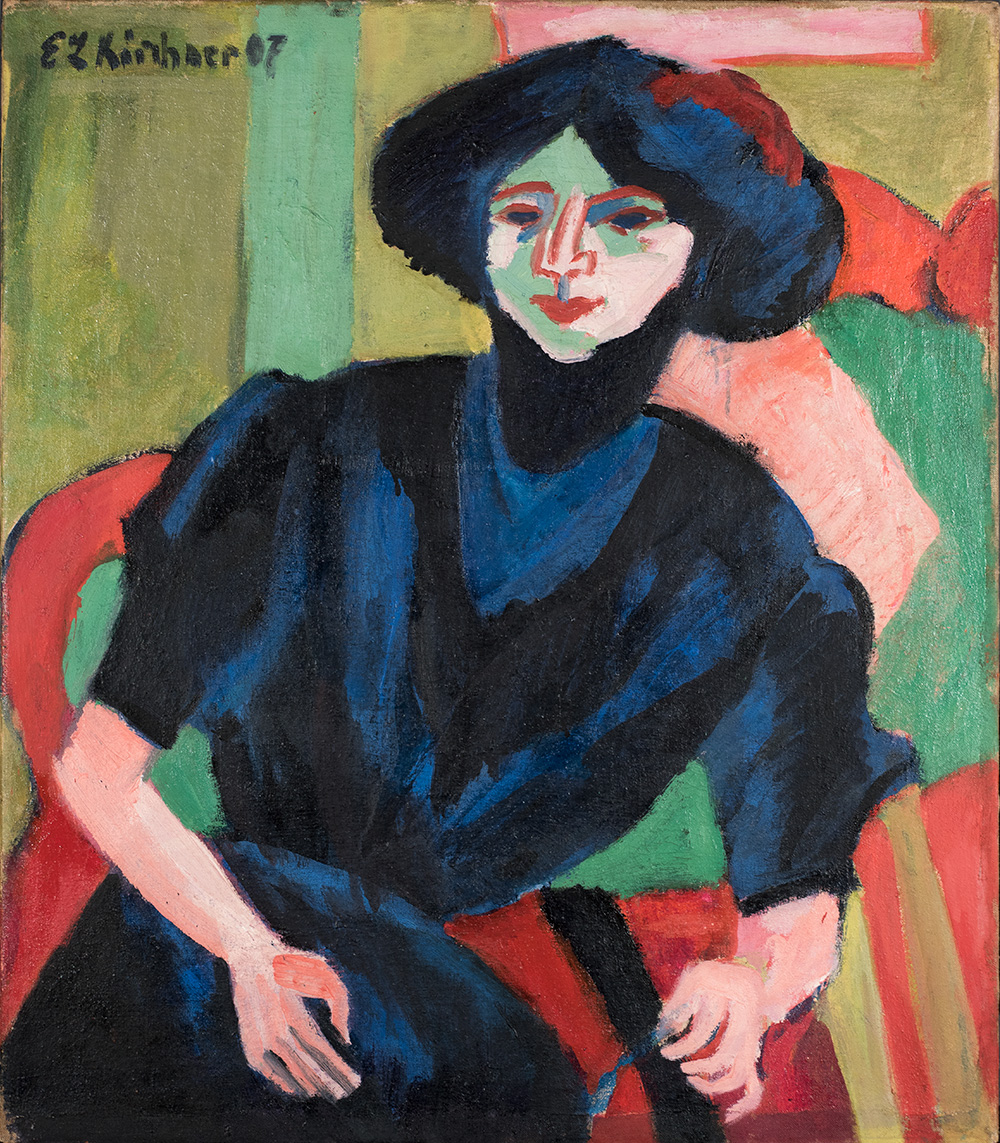
Normal light
Kirchner’s Portrait of Gerti presents one of those exciting examples in technical analysis where the deeper that researcher dives into the material layers, the more questions arise about Kirchner’s process.
The technical images confirm that Kirchner experimented with this painted canvas thoroughly—he changed the sizing format, made changes in the composition, and likely returned at a later date to add more paint, possibly to address condition issues from the inherent vice of his evolving techniques.
Ernst Ludwig Kirchner, German (active Switzerland), 1880–1938; Portrait of Gerti, c.1910–11 (dated 1907); oil on canvas; 31 3/4 x 27 3/4 inches; Saint Louis Art Museum, Given by Sam J. Levin and Audrey L. Levin 26:1992
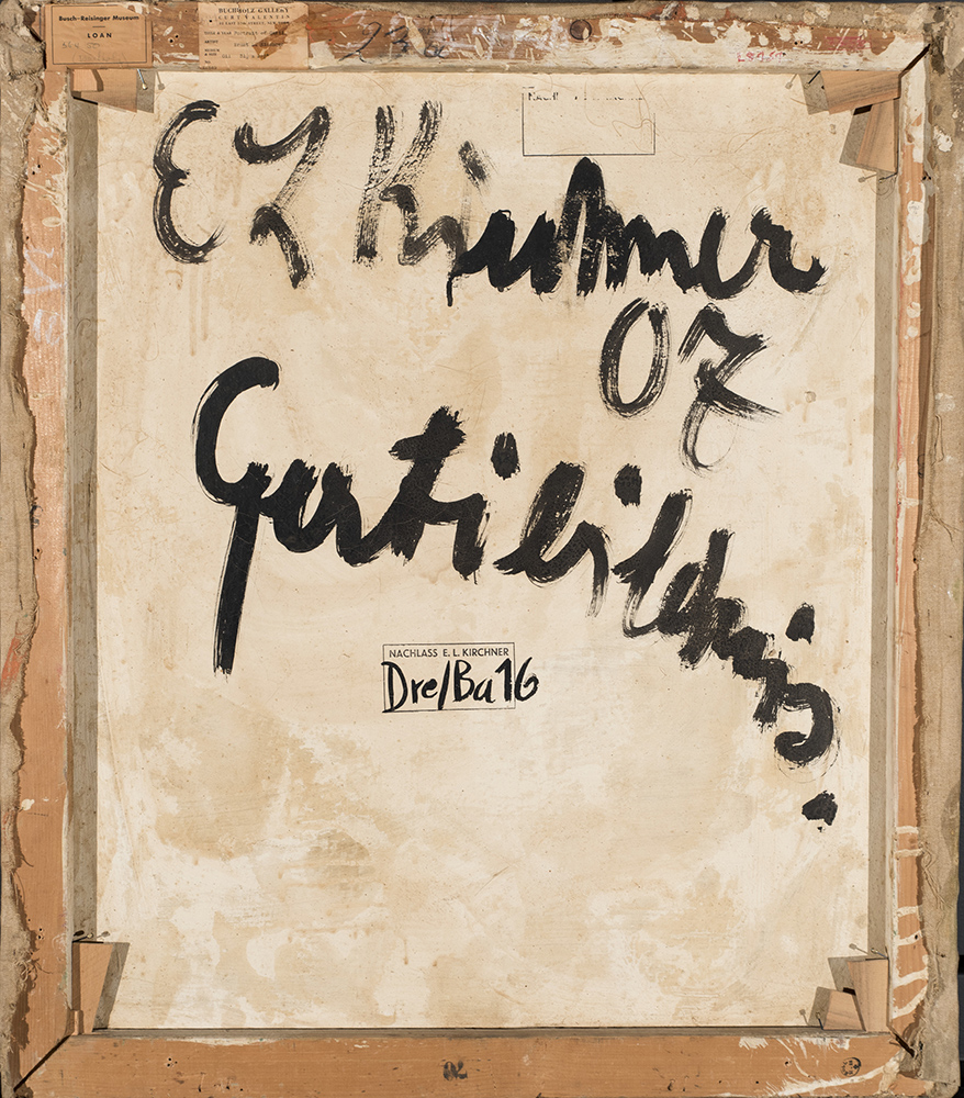
Normal light, back of painting
Kirchner signed, dated, and titled this work “Gertibildnis” in fluid, expressive black script. Kirchner applied a slurry of chalk, glue, and linseed oil to the reverse of the canvas. He brushed on the mixture using broad strokes that dripped and splashed onto the wooden stretcher and canvas—demonstrating that he applied the slurry once he had finished stretching Portrait of Gerti.
This matte mixture is nearly identical to the absorbent ground layer applied on the painting’s front.
Specular light
The high proportion of chalk in Kirchner’s ground layer was designed to absorb oil from the oil paint, and create a matte, velvety surface. Kirchner also was known to add wax to his paints, which softened their sheen. In the specular image, this effect is clearest in the blue and black paints. Shinier areas are likely due to residual conservation materials applied to adhere the separating paint layers. Cracking patterns and losses in the paint layers suggest that the painting has suffered a history of inherent vice, that is, condition issues resulting from incompatible materials and layering techniques.
Note the contrasting strips of missing ground layer located along the bottom and right edges. These areas track where Kirchner expanded the composition—a first clue that he was experimenting with this painting. Instead of applying more ground, he simply covered the exposed raw canvas with paint.

Raking light, cast from right
Light cast from a sharp side angle onto the painting reveals a highly textured surface. Gritty clusters of chalk in his ground layer combine with dried bits of paint—likely scrapings from a well-used paint palette—to create a rough finished texture.
Crests of impasto, especially in the figure’s face and hands, further heighten the effect. Shapes that contrast with the finished composition, such as the hooked formations in the figure’s hand and to the left of her hair within the stripe of chrome green, hint at additional paint layers underneath.
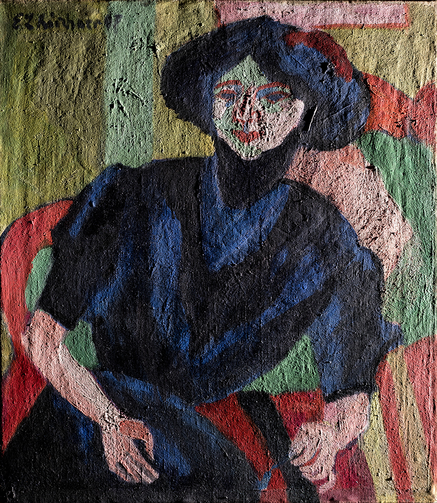
Ultraviolet-induced fluorescent (UVF)
The subtle variations of blue and black in the figure’s dress and hair become vibrantly clear in ultraviolet (UV) light. While the lighter red reads as one hue to the naked eye, an even brighter red peeks from beneath in UV—signifying that Kirchner has matched one with a similar color but chemically different paint tube in what was likely two different campaigns.
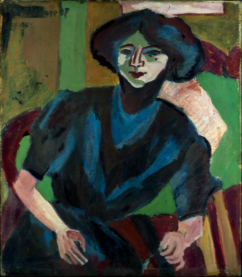
Infrared
Infrared reflectography further showcases the variable brushwork Kirchner has applied to the figure’s dress and hair. Striations made by the shallow sweeping of brush bristles demonstrate how the fluid paint, thinned with solvent, was applied—reinforced by the drip marks running from her coiffed hair.
The green pigments behave differently in the infrared image when compared to the UV image. The chrome green behind Gerti becomes brighter and transparent, revealing the originally larger pink shape underneath.

X-radiograph
When an infrared image indicates changes have been made by the artist, X-radiography is often an excellent complementary technique. The X-radiograph of Portrait of Gerti unexpectedly presented more questions than answers.
The X-radiograph shows the bones of the painting’s wooden stretcher, tacks, and pins quite clearly, since denser and metallic material appears brighter from their X-ray absorption. The swoops of the slurry brushed onto the reverse are also visible, since they were applied quite thickly.
Notably, the composition is scattered with strange, jagged, bright artifacts. Artifacts occasionally line up with the final composition, such as the instance of the figure’s face and the pink shape behind her. Others, such as those above her hand holding a thread and the hooked formation to the left of her hair within the stripe of chrome green, appear foreign. The artifacts suggest concentrations of a metal-dense paint, likely lead, were applied in a lower layer. These areas also corresponded to the contrasting impasto visible in raking light. The mystery of a possible underlying composition warranted a deeper investigation.

Annotated diagram
Since it was suspected that a possible, earlier composition lay beneath Portrait of Gerti, the first task was simply to return to the painting and to look closer. This image maps all the instances where the ground layer or raw canvas appears within the painted boundaries of the final composition. The highlighted areas trace the Portrait of Gerti so faithfully; therefore, any divergent composition was likely placed beneath the applied ground layers. Kirchner, apart from smaller alterations to the pink shape and to the extension of the canvas size, appeared to have followed his design.

Microscopic image of a cross-section
A microscopic sample—smaller than the head of a pin—was carefully extracted from the boundary of the figure’s resting arm and the green shape behind it. This sample was then cast in resin and cut in half to expose the layered structure of applied paint.

Under magnification, the layers of the cross-section are, from top to bottom:
1) green paint (glaze)
2) pink paint
3) blue paint (figure’s dress)
4) ground layer
5) orange-red pigment
The orange-red pigment is likely red lead, which helps explain the strange bright artifacts visible in the X-radiograph due to its ability to strongly absorb X-rays.
Microscopy also helped confirm that the pigment rests underneath the ground layer, applied to raw canvas. What remains a mystery is if the red lead layer mapped out an initial iteration of Gerti or an entirely different composition.
Microscopic detail
Revisiting the painted surface of Portrait of Gerti, the orange-red lead appears to have been thinly painted to sketch the composition, along with a deep blue, likely Prussian blue. These two colors weave over and under each other as they trace the outlines of the present shapes.
In areas where red lead was applied thickly, chemical changes can appear as small blisters in the paint layers. This is due to the formation of lead soaps, where metallic agents in the pigment mix with aging linseed oil in the paint to gradually erupt at the surface. Seen through the lens of a microscope, one such blister in the paint layer, located in the green stripe to the left of the figure’s head, shows red lead, rimmed with a layer of white ground layer, erupting through the green paint.

Additional Objects
View all objects in the technical imaging suite to reveal hidden details and examine underlying material layers.

-
Glossary
Back (Verso)
The back of a painting provides a wealth of information about its structure. Changes made to the canvas, such as visible repairs, reveal past damage or vulnerabilities. Markings, labels, and inscriptions often tell new stories of the painting’s past, including original titles, previous owners, and exhibition histories.
Raking Light
Raking light, directed from a side angle, casts surface textures into strong relief. This technique highlights details such as thick brushstrokes, canvas weave, or cracks in the paint layers.
Specular Light
Light cast directly onto the painting’s surface at a 90-degree angle highlights glossy or matte qualities. This technique helps determine if a varnish is present and quickly exposes variations of sheen in different paints.
Infrared Reflectography
Infrared reflectography (IRR) uses a specially adapted camera to detect infrared energy. When an image of a painting is captured using infrared light, some pigments appear transparent, which enables the viewer to see through overlying layers of paint. This technique often reveals hidden details, such as preparatory drawings by the artist or parts of the composition the artist painted over.
Ultraviolet Radiation
Ultraviolet (UV) light sources cause certain materials to absorb and re-emit colorful light, known as fluorescence. The fluorescence of some materials can identify certain pigments, such as zinc white, which produces a vibrant glow. Conservators can also detect previous treatments, which may appear dark purple.
X-radiography
X-radiography uses an X-ray source and sensitive film to produce an image of the painting’s composite layers. Materials transparent to X-rays appear dark in the images, while X-ray–absorbing materials like lead white and metal hardware appear white. Artist’s changes and, in some exciting instances, completely different, earlier compositions may be unveiled.